Copyright
©2006 Baishideng Publishing Group Co.
World J Gastroenterol. Aug 14, 2006; 12(30): 4914-4917
Published online Aug 14, 2006. doi: 10.3748/wjg.v12.i30.4914
Published online Aug 14, 2006. doi: 10.3748/wjg.v12.i30.4914
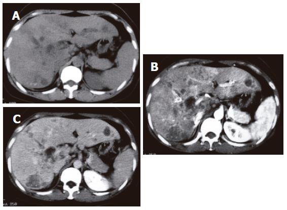
Figure 2 Abdominal computed tomography (CT).
A: Plain CT images show intrahepatic ductal dilatation, micro-calcifications, and multiple hypodense lesions in the liver; B: Contrast CT image (early phase) shows clearly delineated hypodense lesions; C: Late phase CT image shows slightly enhanced hypodense lesions.
- Citation: Iwai T, Kida M, Kida Y, Shikama N, Shibuya A, Saigenji K. Biliary tuberculosis causing cicatricial stenosis after oral anti-tuberculosis therapy. World J Gastroenterol 2006; 12(30): 4914-4917
- URL: https://www.wjgnet.com/1007-9327/full/v12/i30/4914.htm
- DOI: https://dx.doi.org/10.3748/wjg.v12.i30.4914


