Copyright
©2006 Baishideng Publishing Group Co.
World J Gastroenterol. Jan 21, 2006; 12(3): 426-430
Published online Jan 21, 2006. doi: 10.3748/wjg.v12.i3.426
Published online Jan 21, 2006. doi: 10.3748/wjg.v12.i3.426
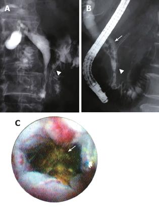
Figure 2 No bile duct stone was observed immediately after EMS in patient number 7 (A), however, stones were noted 6.
1 years later (arrow), and they were successfully removed using a basket wire (B). Peroral cholangioscopy revealed the common bile duct distal to the stone (arrow) was patent and the hyperplastic change was not conspicuous (C). Arrow head indicates the pancreatic duct stent.
- Citation: Yamaguchi T, Ishihara T, Seza K, Nakagawa A, Sudo K, Tawada K, Kouzu T, Saisho H. Long-term outcome of endoscopic metallic stenting for benign biliary stenosis associated with chronic pancreatitis. World J Gastroenterol 2006; 12(3): 426-430
- URL: https://www.wjgnet.com/1007-9327/full/v12/i3/426.htm
- DOI: https://dx.doi.org/10.3748/wjg.v12.i3.426


