Copyright
©2006 Baishideng Publishing Group Co.
World J Gastroenterol. Jan 21, 2006; 12(3): 363-371
Published online Jan 21, 2006. doi: 10.3748/wjg.v12.i3.363
Published online Jan 21, 2006. doi: 10.3748/wjg.v12.i3.363
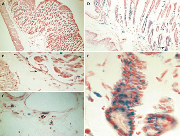
Figure 4 Engraftment of donor-derived ROSA-26 marrow by x-gal staining.
A: Mice transplanted with ROSA 26 marrow and infected with H. felis for 4 wk had donor-derived leukocytes (blue) infiltrating the gastric mucosa, and no engraftment into gland structures. B and C: A higher power view reveals myocytes and myofibroblasts in the submucosal tissue adjacent to vascular structures (arrows). D: After 30 wk of infection, marked architectural distortion is seen with antralization and appearance of metaplastic glands. Entire gland structures are derived from donor marrow (blue staining). Gland shown in panel D (arrow) is shown at higher power in E.
- Citation: Li HC, Stoicov C, Rogers AB, Houghton J. Stem cells and cancer: Evidence for bone marrow stem cells in epithelial cancers. World J Gastroenterol 2006; 12(3): 363-371
- URL: https://www.wjgnet.com/1007-9327/full/v12/i3/363.htm
- DOI: https://dx.doi.org/10.3748/wjg.v12.i3.363


