Copyright
©2006 Baishideng Publishing Group Co.
World J Gastroenterol. Jul 28, 2006; 12(28): 4599-4601
Published online Jul 28, 2006. doi: 10.3748/wjg.v12.i28.4599
Published online Jul 28, 2006. doi: 10.3748/wjg.v12.i28.4599
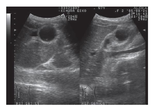
Figure 1 Abdominal ultrasonography revealed an abnormally large floating gallbladder without gallstones, and a thickened gallbladder wall.
- Citation: Matsuhashi N, Satake S, Yawata K, Asakawa E, Mizoguchi T, Kanematsu M, Kondo H, Yasuda I, Nonaka K, Tanaka C, Misao A, Ogura S. Volvulus of the gall bladder diagnosed by ultrasonography, computed tomography, coronal magnetic resonance imaging and magnetic resonance cholangio-pancreatography. World J Gastroenterol 2006; 12(28): 4599-4601
- URL: https://www.wjgnet.com/1007-9327/full/v12/i28/4599.htm
- DOI: https://dx.doi.org/10.3748/wjg.v12.i28.4599


