Copyright
©2006 Baishideng Publishing Group Co.
World J Gastroenterol. Jul 28, 2006; 12(28): 4588-4592
Published online Jul 28, 2006. doi: 10.3748/wjg.v12.i28.4588
Published online Jul 28, 2006. doi: 10.3748/wjg.v12.i28.4588
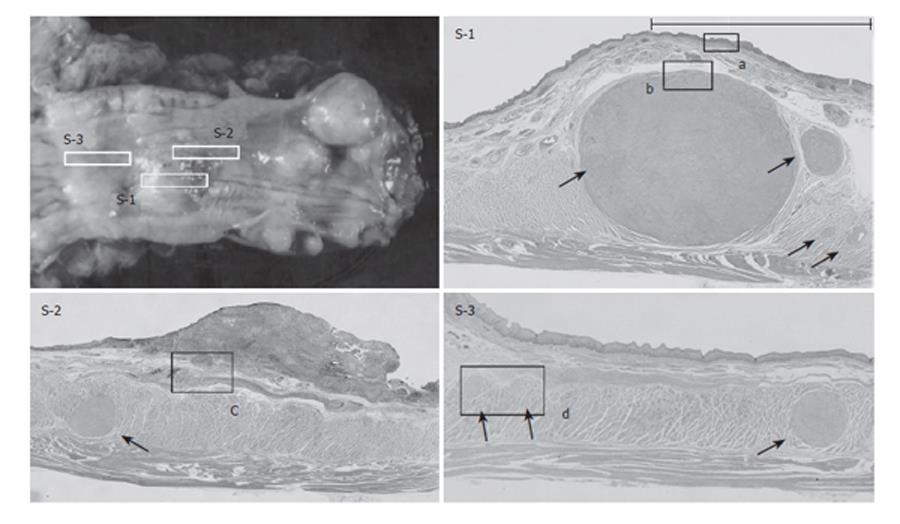
Figure 4 Photomicrograph of cross-sections (squares) of the resected specimen.
Leiomyoma tissue is located in all sections (arrows). S-1: CIS overlies the leiomyoma. The region of CIS extends to the oral side of the leiomyoma (bar). S-2: SCC invades the submucosal layer in a very narrow region. S-3: A section without abnormal macroscopic findings. Multiple small leiomyomas are observed only by microscopic examination.
- Citation: Iwaya T, Maesawa C, Uesugi N, Kimura T, Ikeda K, Kimura Y, Mitomo S, Ishida K, Sato N, Wakabayashi G. Coexistence of esophageal superficial carcinoma and multiple leiomyomas: A case report. World J Gastroenterol 2006; 12(28): 4588-4592
- URL: https://www.wjgnet.com/1007-9327/full/v12/i28/4588.htm
- DOI: https://dx.doi.org/10.3748/wjg.v12.i28.4588


