Copyright
©2006 Baishideng Publishing Group Co.
World J Gastroenterol. Jul 28, 2006; 12(28): 4524-4528
Published online Jul 28, 2006. doi: 10.3748/wjg.v12.i28.4524
Published online Jul 28, 2006. doi: 10.3748/wjg.v12.i28.4524
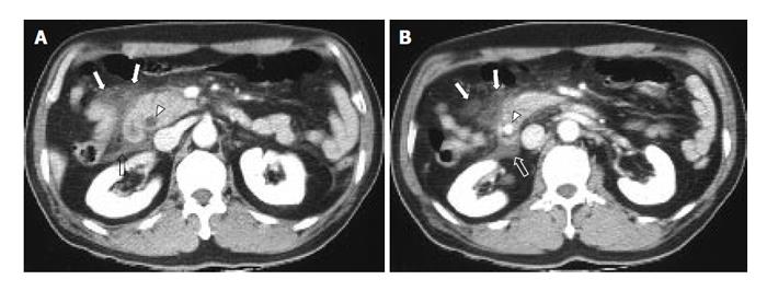
Figure 2 A 61-year old man with a clinical diagnosis of gallstone pancreatitis due to distal common bile duct stone.
A and B: CT scans showing the peripancreatic infiltration or fluid collection predominantly in the right abdomen: score 1 infiltration in RP compartment (arrows), score 2 fluid collection in RSR compartment (open arrows). Distal common bile duct stone with bile duct dilatation also can be seen (arrowheads).
- Citation: Kim YS, Kim Y, Kim SK, Rhim H. Computed tomographic differentiation between alcoholic and gallstone pancreatitis: Significance of distribution of infiltration or fluid collection. World J Gastroenterol 2006; 12(28): 4524-4528
- URL: https://www.wjgnet.com/1007-9327/full/v12/i28/4524.htm
- DOI: https://dx.doi.org/10.3748/wjg.v12.i28.4524


