Copyright
©2006 Baishideng Publishing Group Co.
World J Gastroenterol. Jul 21, 2006; 12(27): 4281-4295
Published online Jul 21, 2006. doi: 10.3748/wjg.v12.i27.4281
Published online Jul 21, 2006. doi: 10.3748/wjg.v12.i27.4281
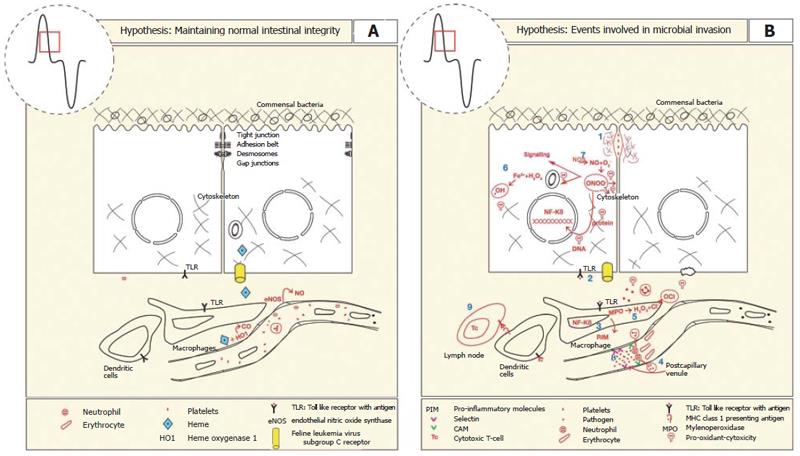
Figure 4 A: The intestinal mucosal barrier is maintained by a series of lateral membrane specialisations near the apical pole of the epithelial cell.
It comprises tight junctions, adhesion belts, desmosomes and gap junctions that prevent the movement of pathogens across the epithelial monolayer. Constitutive expression and activities of endothelial nitric oxide synthase (eNOS) and heme oxygenase 1 (HO-1) is important for maintaining adequate blood flow, anti-inflammatory, anti-thromobotic (-) and anti-apoptotic effects on endothelium, neutrophils, platelets, and enterocytes, respectively. HO-1 activity produces the antioxidant bilirubin to limit oxidative damage; B: The loss of mucosal integrity results in the translocation of pathogens and establishment of an inflammatory response by the following series of events. (1) Initiation of synthesis of proteases by bacteria erode tight junction complexes between epithelial cells. (2) Binding of bacterial motifs activates toll like receptors, initiating the NF-kB pathway. (3) Increased expression of pro-inflammatory cytokines, chemokines and endothelial cell surface adhesion molecules. (4) Leukocytes extravasate and increased permeability of capillaries increases fluid accumulation (5) Phagocytic cells produce myeloperoxidase which combines with peroxide to form hypochlorous acid that damages pathogen and host systems alike. (6) Peroxide produced by enterocytes in combination with ferrous iron can produce superoxide anions that damage lipids, DNA and proteins. (7) NO is produced at high concentrations that combines with peroxide to form the pro-oxidant peroxynitrite. (8) Platelets also bind to the endothelial surface to induce hemostasis. (9) Presentation of antigens by dendritic cells via major histocompatibility class 1 to cytotoxic T-cells leads to antibody presentation and destruction of infected epithelial cells.
- Citation: Oates PS, West AR. Heme in intestinal epithelial cell turnover, differentiation, detoxification, inflammation, carcinogenesis, absorption and motility. World J Gastroenterol 2006; 12(27): 4281-4295
- URL: https://www.wjgnet.com/1007-9327/full/v12/i27/4281.htm
- DOI: https://dx.doi.org/10.3748/wjg.v12.i27.4281


