Copyright
©2006 Baishideng Publishing Group Co.
World J Gastroenterol. Jul 7, 2006; 12(25): 4104-4105
Published online Jul 7, 2006. doi: 10.3748/wjg.v12.i25.4104
Published online Jul 7, 2006. doi: 10.3748/wjg.v12.i25.4104
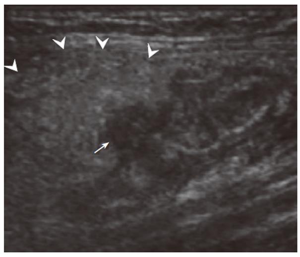
Figure 1 Longitudinal view of the appendix.
A hyperechoic mass (arrowheads) including hypoechoic lateral pouch like lesion (arrow) is observed. Hypo lesion is appendiceal diverticula, and hyperechoic mass is inflamed adipose tissue (= mesoappendix).
- Citation: Kubota T, Omori T, Yamamoto J, Nagai M, Tamaki S, Sasaki K. Sonographic findings of acute appendiceal diverticulitis. World J Gastroenterol 2006; 12(25): 4104-4105
- URL: https://www.wjgnet.com/1007-9327/full/v12/i25/4104.htm
- DOI: https://dx.doi.org/10.3748/wjg.v12.i25.4104


