Copyright
©2006 Baishideng Publishing Group Co.
World J Gastroenterol. Jul 7, 2006; 12(25): 4038-4043
Published online Jul 7, 2006. doi: 10.3748/wjg.v12.i25.4038
Published online Jul 7, 2006. doi: 10.3748/wjg.v12.i25.4038
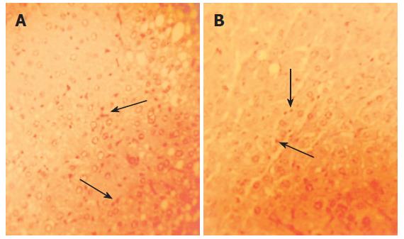
Figure 3 Representative examples of orcein staining of HBV-Tg liver (× 400).
HBsAg was seen as nigger-brown granules before drug administration (A) and after 10 wk of therapy by Sp (B). However, a decrease in the signals for HBsAg was seen in (B) compared with that in (A).
-
Citation: Wang R, Du ZL, Duan WJ, Zhang X, Zeng FL, Wan XX. Antiviral treatment of hepatitis B virus-transgenic mice by a marine organism,
Styela plicata . World J Gastroenterol 2006; 12(25): 4038-4043 - URL: https://www.wjgnet.com/1007-9327/full/v12/i25/4038.htm
- DOI: https://dx.doi.org/10.3748/wjg.v12.i25.4038


