Copyright
©2006 Baishideng Publishing Group Co.
World J Gastroenterol. Jun 28, 2006; 12(24): 3821-3828
Published online Jun 28, 2006. doi: 10.3748/wjg.v12.i24.3821
Published online Jun 28, 2006. doi: 10.3748/wjg.v12.i24.3821
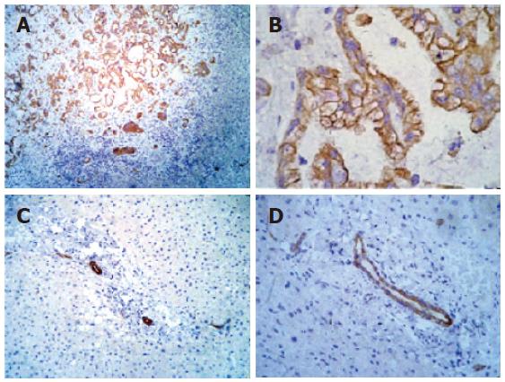
Figure 1 Immunohistochemical analysis of IGF-IR in liver tissue.
A: Positive control cholangiocarcinoma tissue showing strongly positive staining for IGF-IR in neoplastic ducts (-100 ×); B: positive control cholangiocarcinoma tissue showing strongly positive staining for IGF-IR in neoplastic ducts (-200 ×); C: normal liver tissue showing IGF-IR immunoreactivity in ductal cells (-100 ×); D: normal liver tissue showing IGF-IR immunoreactivity in ductal cells (-200 ×).
- Citation: Stefano JT, Corrêa-Giannella ML, Ribeiro CMF, Alves VAF, Massarollo PCB, Machado MCC, Giannella-Neto D. Increased hepatic expression of insulin-like growth factor-I receptor in chronic hepatitis C. World J Gastroenterol 2006; 12(24): 3821-3828
- URL: https://www.wjgnet.com/1007-9327/full/v12/i24/3821.htm
- DOI: https://dx.doi.org/10.3748/wjg.v12.i24.3821


