Copyright
©2006 Baishideng Publishing Group Co.
World J Gastroenterol. Jun 21, 2006; 12(23): 3729-3735
Published online Jun 21, 2006. doi: 10.3748/wjg.v12.i23.3729
Published online Jun 21, 2006. doi: 10.3748/wjg.v12.i23.3729
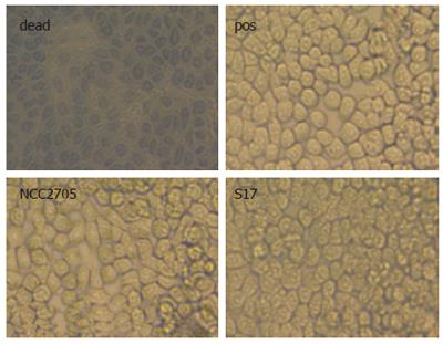
Figure 3 Microscopic examination of cellular viability.
Trypan blue exclusion was monitored in HT-29 clone 34 cells challenged with LPS + HM for 16 after pre-incubation with bifidobacteria (B. longum NCC2705 or B. bifidum S17; moi = 100). Positive control (pos) was cells incubated with LPS + HM without bacterial pre-treatment. As a control for trypan blue staining cells were incubated for 16 h in cell culture medium at pH 4 (dead).
- Citation: Riedel CU, Foata F, Philippe D, Adolfsson O, Eikmanns BJ, Blum S. Anti-inflammatory effects of bifidobacteria by inhibition of LPS-induced NF-κB activation. World J Gastroenterol 2006; 12(23): 3729-3735
- URL: https://www.wjgnet.com/1007-9327/full/v12/i23/3729.htm
- DOI: https://dx.doi.org/10.3748/wjg.v12.i23.3729


