Copyright
©2006 Baishideng Publishing Group Co.
World J Gastroenterol. Jun 14, 2006; 12(22): 3546-3552
Published online Jun 14, 2006. doi: 10.3748/wjg.v12.i22.3546
Published online Jun 14, 2006. doi: 10.3748/wjg.v12.i22.3546
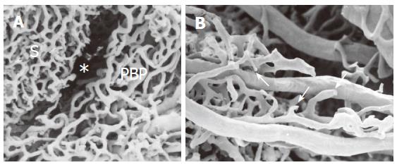
Figure 3 SEMvcc of BDL rat liver (orig.
magn., A: 40×; B: 60×). (A) proliferated PBP run at the periphery of the lobule separated from the sinusoidal network (S) by an empty space (*) which corresponds to proliferating connective tissue. (B) not completely developed PBP with typical neovascular organization with dead end vessels (arrows).
- Citation: Gaudio E, Franchitto A, Pannarale L, Carpino G, Alpini G, Francis H, Glaser S, Alvaro D, Onori P. Cholangiocytes and blood supply. World J Gastroenterol 2006; 12(22): 3546-3552
- URL: https://www.wjgnet.com/1007-9327/full/v12/i22/3546.htm
- DOI: https://dx.doi.org/10.3748/wjg.v12.i22.3546


