Copyright
©2006 Baishideng Publishing Group Co.
World J Gastroenterol. Jun 14, 2006; 12(22): 3512-3522
Published online Jun 14, 2006. doi: 10.3748/wjg.v12.i22.3512
Published online Jun 14, 2006. doi: 10.3748/wjg.v12.i22.3512
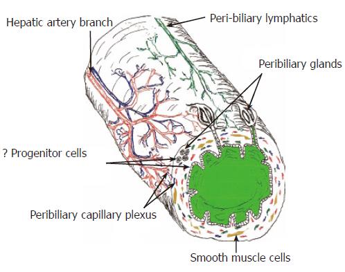
Figure 1 Diagram of the extra-hepatic and large intra-hepatic bile ducts highlighting some important anatomic and physiologic considerations that can potentially impact wound healing.
Except for the peribiliary glands and some features of the BEC, small intra-hepatic bile ducts have a similar anatomy. All of the bile ducts, including the small intra-hepatic bile ducts are supplied only by the hepatic artery and the peribiliary vascular plexus, shown in red. All bile ducts also contain either smooth muscle cells and/or facultative myofibroblasts in their wall. Deep wounds to the biliary tree result in activation and/or transformation of myofibroblasts that greatly increase the risk of wound contraction, fibrosis, and stricture formation.
- Citation: Demetris AJ, III JGL, Specht S, Nozaki I. Biliary wound healing, ductular reactions, and IL-6/gp130 signaling in the development of liver disease. World J Gastroenterol 2006; 12(22): 3512-3522
- URL: https://www.wjgnet.com/1007-9327/full/v12/i22/3512.htm
- DOI: https://dx.doi.org/10.3748/wjg.v12.i22.3512


