Copyright
©2006 Baishideng Publishing Group Co.
World J Gastroenterol. Jun 14, 2006; 12(22): 3461-3465
Published online Jun 14, 2006. doi: 10.3748/wjg.v12.i22.3461
Published online Jun 14, 2006. doi: 10.3748/wjg.v12.i22.3461
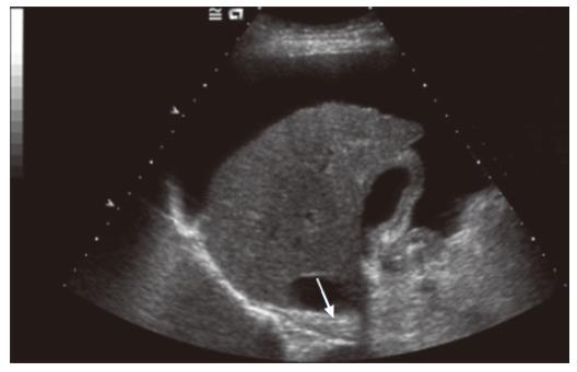
Figure 3 Ultrasound image of a typical cirrhotic liver with a shrunken right lobe, a nodular surface (white arrow), surrounding ascites and a heterogeneous echotexture.
- Citation: Grier S, Lim AK, Patel N, Cobbold JF, Thomas HC, Cox IJ, Taylor-Robinson SD. Role of microbubble ultrasound contrast agents in the non-invasive assessment of chronic hepatitis C-related liver disease. World J Gastroenterol 2006; 12(22): 3461-3465
- URL: https://www.wjgnet.com/1007-9327/full/v12/i22/3461.htm
- DOI: https://dx.doi.org/10.3748/wjg.v12.i22.3461


