Copyright
©2006 Baishideng Publishing Group Co.
World J Gastroenterol. May 28, 2006; 12(20): 3265-3270
Published online May 28, 2006. doi: 10.3748/wjg.v12.i20.3265
Published online May 28, 2006. doi: 10.3748/wjg.v12.i20.3265
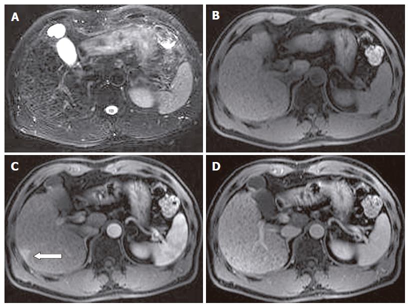
Figure 2 HPD in a 56-year-old man with liver cirrhosis.
A: Fat-saturated breath-hold turbo FSE T2-weighted transverse MR scan shows multiple punctuate hypointensity cirrhotic nodules and surrounding reticulated fibrotic liver parenchyma, without any obvious hyperintense tumoral lesions; B: pre- contrast administration transverse spoiled gradient-echo T1-weighted MR image (118/4.1; flip angle, 80°) shows no obvious hypointense tumoral lesions observed; C: transverse contrast-enhanced T1-weighted MR image obtained during HAP shows a subcapsular small patchy hyperintense tumor-like lesion (pseudolesion) (open arrow); D: transverse contrast-enhanced T1-weighted MR image obtained during PVP shows no lesion in that area.
- Citation: Tian JL, Zhang JS. Hepatic perfusion disorders: Etiopathogenesis and related diseases. World J Gastroenterol 2006; 12(20): 3265-3270
- URL: https://www.wjgnet.com/1007-9327/full/v12/i20/3265.htm
- DOI: https://dx.doi.org/10.3748/wjg.v12.i20.3265


