Copyright
©2006 Baishideng Publishing Group Co.
World J Gastroenterol. May 28, 2006; 12(20): 3139-3145
Published online May 28, 2006. doi: 10.3748/wjg.v12.i20.3139
Published online May 28, 2006. doi: 10.3748/wjg.v12.i20.3139
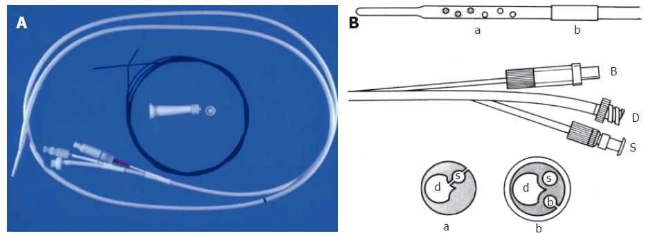
Figure 8 A: Image of MDEC decompression and enteroclysis tube with stiffening wire that helps maneuvering the tube into the proximal jejunum; B: Line diagram of the tube.
B = balloon port, D = drainage port, S = sump port (helps prevent the tube from becoming obstructed by debris). The inset figures show the cross sectional appearances of the tube at positions a and b.
- Citation: Maglinte DD, Sandrasegaran K, Tann M. Advances in alimentary tract imaging. World J Gastroenterol 2006; 12(20): 3139-3145
- URL: https://www.wjgnet.com/1007-9327/full/v12/i20/3139.htm
- DOI: https://dx.doi.org/10.3748/wjg.v12.i20.3139


