Copyright
©2006 Baishideng Publishing Group Co.
World J Gastroenterol. May 28, 2006; 12(20): 3139-3145
Published online May 28, 2006. doi: 10.3748/wjg.v12.i20.3139
Published online May 28, 2006. doi: 10.3748/wjg.v12.i20.3139
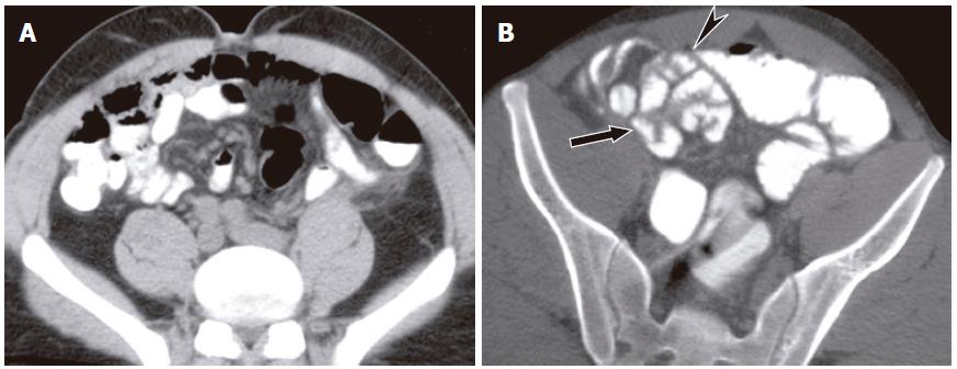
Figure 7 Fifty-four years old with prior hysterectomy presented with abdominal bloating and nausea.
A: Conventional CT did not show distended bowel or transition point and was interpreted as normal; B: CT enteroclysis performed a day later shows distended small bowel loop with beaking (arrowhead) adjacent to anterior parietal peritoneum. Distal small bowel loops were nondistended (arrow). The appearances were of low grade small bowel obstruction due to adhesions.
- Citation: Maglinte DD, Sandrasegaran K, Tann M. Advances in alimentary tract imaging. World J Gastroenterol 2006; 12(20): 3139-3145
- URL: https://www.wjgnet.com/1007-9327/full/v12/i20/3139.htm
- DOI: https://dx.doi.org/10.3748/wjg.v12.i20.3139


