Copyright
©2006 Baishideng Publishing Group Co.
World J Gastroenterol. May 28, 2006; 12(20): 3139-3145
Published online May 28, 2006. doi: 10.3748/wjg.v12.i20.3139
Published online May 28, 2006. doi: 10.3748/wjg.v12.i20.3139
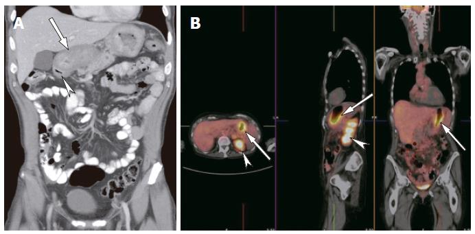
Figure 6 Sixty-nine years old male with dyspepsia.
A: Coronal reformat showed diffuse thickening of the gastric wall (arrow). The duodenal cap was normal (arrowhead); B: Fused PET-CT images showed hypermetabolic activity of the gastric wall indicating cancer. No other foci of abnormal uptake were identified. Note the normal increased activity in the renal collecting systems (arrowheads) due to excretion of radiopharmaceutical.
- Citation: Maglinte DD, Sandrasegaran K, Tann M. Advances in alimentary tract imaging. World J Gastroenterol 2006; 12(20): 3139-3145
- URL: https://www.wjgnet.com/1007-9327/full/v12/i20/3139.htm
- DOI: https://dx.doi.org/10.3748/wjg.v12.i20.3139


