Copyright
©2006 Baishideng Publishing Group Co.
World J Gastroenterol. May 28, 2006; 12(20): 3139-3145
Published online May 28, 2006. doi: 10.3748/wjg.v12.i20.3139
Published online May 28, 2006. doi: 10.3748/wjg.v12.i20.3139
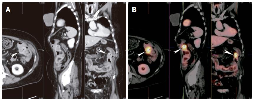
Figure 5 Thirty-nine years old female with prior sigmoid colectomy for cancer, presented with rising CA-125.
A: Axial, coronal and sagittal images of CT raised concern for possible mass adjacent to the splenic flexure (arrowheads); B: Fused images of PET and CT showed hypermetabolic focus (arrowheads) in the mesocolon adjacent to splenic flexure, indicating recurrent tumor. No other sites of tumor were found. While PET is sensitive for recurrent disease it has poor spatial resolution and it would have been difficult to determine if the recurrence was in the colon, mesentery or adjacent spleen. Fusion of PET and CT images allowed accurate anatomical localization.
- Citation: Maglinte DD, Sandrasegaran K, Tann M. Advances in alimentary tract imaging. World J Gastroenterol 2006; 12(20): 3139-3145
- URL: https://www.wjgnet.com/1007-9327/full/v12/i20/3139.htm
- DOI: https://dx.doi.org/10.3748/wjg.v12.i20.3139


