Copyright
©2006 Baishideng Publishing Group Co.
World J Gastroenterol. May 28, 2006; 12(20): 3139-3145
Published online May 28, 2006. doi: 10.3748/wjg.v12.i20.3139
Published online May 28, 2006. doi: 10.3748/wjg.v12.i20.3139
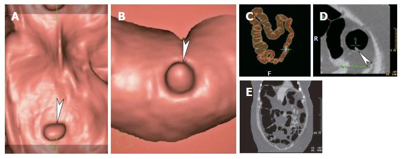
Figure 4 Virtual colonoscopy images in 62 years old female with incomplete optical colonoscopy.
A: Dissection or fillet view; B: Endoluminal fly-through view; C: Transparent localizing view; D: Two dimensional axial view; E: Two dimensional coronal view. At our institution the initial read is the 3 dimensional fillet and endoluminal views in antegrade and retrograde fashion. Sites of concern are correlated with axial or coronal two dimensional views. The entire interpretation including assessment of extracolonic structures takes about 30 min. Note the 6 mm polyp (arrowhead). Its location was confirmed as distal descending colon on image C.
- Citation: Maglinte DD, Sandrasegaran K, Tann M. Advances in alimentary tract imaging. World J Gastroenterol 2006; 12(20): 3139-3145
- URL: https://www.wjgnet.com/1007-9327/full/v12/i20/3139.htm
- DOI: https://dx.doi.org/10.3748/wjg.v12.i20.3139


