Copyright
©2006 Baishideng Publishing Group Co.
World J Gastroenterol. May 28, 2006; 12(20): 3139-3145
Published online May 28, 2006. doi: 10.3748/wjg.v12.i20.3139
Published online May 28, 2006. doi: 10.3748/wjg.v12.i20.3139
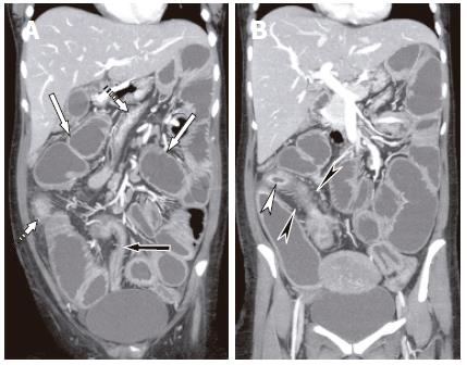
Figure 2 Abdominal pain and fever in 23 years old female.
A, B: Coronal reformats with neutral enteral contrast (water) showed dilated proximal loops of small bowel (white solid arrows), and nondistended colon (dashed arrows). A segment of distal ileum (black arrow, image A) showed stenosis with mucosal enhancement, consistent with inflammatory stricture causing small bowel obstruction. A second noncontiguous segment of terminal ileum showed intense mucosal enhancement (white arrowhead, image B) and comb sign due to mesenteric hypervascularity (black arrowhead, image B). The appearances were consistent with acute Crohn’s disease. The diagnosis of wall enhancement and thickening is made easier by using water as oral contrast. The coronal perspective also allows easier evaluation of length of involved segments.
- Citation: Maglinte DD, Sandrasegaran K, Tann M. Advances in alimentary tract imaging. World J Gastroenterol 2006; 12(20): 3139-3145
- URL: https://www.wjgnet.com/1007-9327/full/v12/i20/3139.htm
- DOI: https://dx.doi.org/10.3748/wjg.v12.i20.3139


