Copyright
©2006 Baishideng Publishing Group Co.
World J Gastroenterol. Jan 14, 2006; 12(2): 298-301
Published online Jan 14, 2006. doi: 10.3748/wjg.v12.i2.298
Published online Jan 14, 2006. doi: 10.3748/wjg.v12.i2.298
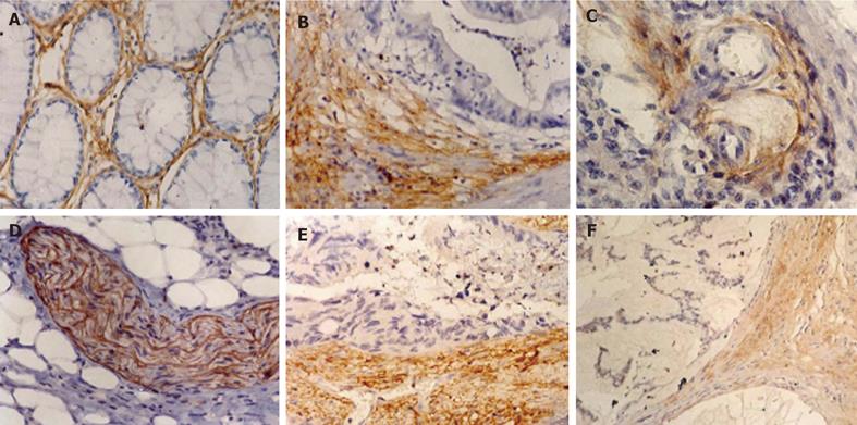
Figure 1 PINCH immunohistochemical staining in normal mucosa (A), adenocarcinoma (B), tunica adventitia of blood vessel (C), peripheral nerve fascicles (D) and stronger expression of PINCH at the invasion edge than in the inner area(E), and at the invasion edge of mucinous carcinoma (F).
- Citation: Zhao ZR, Zhang ZY, Cui DS, Jiang L, Zhang HJ, Wang MW, Sun XF. Particularly interesting new cysteine-histidine rich protein expression in colorectal adenocarcinomas. World J Gastroenterol 2006; 12(2): 298-301
- URL: https://www.wjgnet.com/1007-9327/full/v12/i2/298.htm
- DOI: https://dx.doi.org/10.3748/wjg.v12.i2.298


