Copyright
©2006 Baishideng Publishing Group Co.
World J Gastroenterol. Jan 14, 2006; 12(2): 234-239
Published online Jan 14, 2006. doi: 10.3748/wjg.v12.i2.234
Published online Jan 14, 2006. doi: 10.3748/wjg.v12.i2.234
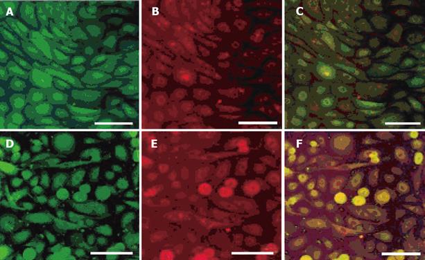
Figure 1 Expression of pepsin, MUC5AC and proton-pump in cultured gastric epithelial cells observed by double immunolabelling.
A confocal microscopic image demonstrates the FITC-positive anti-pepsin-positive cells (A) and Texas Red-positive anti-MUC5AC-positive cells (B) in the same area. The anti-pepsin-positive cells, which co-express MUC5AC, are indicated yellow (C). A confocal microscopic image also demonstrates the FITC-positive anti-pepsin-positive cells (D) and Texas Red-positive anti-proton pump-positive cells (E) in the same area. The anti-pepsin-positive cells which co-express proton pump are indicated yellow (F).Scale bars indicate 100 μm.
- Citation: Itoh K, Sawasaki Y, Takeuchi K, Kato S, Imai N, Kato Y, Shibata N, Kobayashi M, Moriguchi Y, Higuchi M, Ishihata F, Sudoh Y, Miura S. Erythropoietin -induced proliferation of gastric mucosal cells. World J Gastroenterol 2006; 12(2): 234-239
- URL: https://www.wjgnet.com/1007-9327/full/v12/i2/234.htm
- DOI: https://dx.doi.org/10.3748/wjg.v12.i2.234


