Copyright
©2006 Baishideng Publishing Group Co.
World J Gastroenterol. Jan 14, 2006; 12(2): 228-233
Published online Jan 14, 2006. doi: 10.3748/wjg.v12.i2.228
Published online Jan 14, 2006. doi: 10.3748/wjg.v12.i2.228
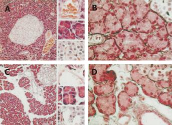
Figure 1 Pancreatic tissues from sham and BDL-60 rats were stained with hematoxylin and eosin (panels A and C) and Gomori (panels B and D).
In panels A and C, magnification is 100x and 400x (upper: small vessel; middle: acini; lower: Langerhans islet) and in panels B and D, magnification is 400x. The extracellular matrix was enlarged in pancreas isolated from BDL-60 rats and most of the enlarged extracellular matrix consisted of edema. No increase in reticulin fibers was observed with the Gomori technique. n = 3 in each group.
- Citation: Frossard JL, Quadri R, Hadengue A, Morel P, Pastor CM. Endothelial nitric oxide synthase regulation is altered in pancreas from cirrhotic rats. World J Gastroenterol 2006; 12(2): 228-233
- URL: https://www.wjgnet.com/1007-9327/full/v12/i2/228.htm
- DOI: https://dx.doi.org/10.3748/wjg.v12.i2.228


