Copyright
©2006 Baishideng Publishing Group Co.
World J Gastroenterol. May 21, 2006; 12(19): 3101-3104
Published online May 21, 2006. doi: 10.3748/wjg.v12.i19.3101
Published online May 21, 2006. doi: 10.3748/wjg.v12.i19.3101
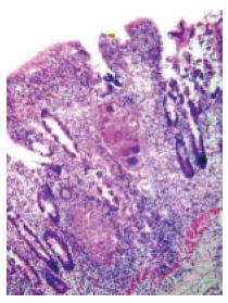
Figure 6 Histological examination of the biopsies obtained from the area of nodularity and ulceration in the terminal ileum showing noncaseating granulomas consisting of Langhans’ giant cells and epithelioid cells.
Note that a cuff of lymphocytes surrounds the granuloma (HE ×80).
- Citation: Misra S, Dwivedi M, Misra V. Ileoscopy in 39 hematochezia patients with normal colonoscopy. World J Gastroenterol 2006; 12(19): 3101-3104
- URL: https://www.wjgnet.com/1007-9327/full/v12/i19/3101.htm
- DOI: https://dx.doi.org/10.3748/wjg.v12.i19.3101


