Copyright
©2006 Baishideng Publishing Group Co.
World J Gastroenterol. May 21, 2006; 12(19): 3020-3025
Published online May 21, 2006. doi: 10.3748/wjg.v12.i19.3020
Published online May 21, 2006. doi: 10.3748/wjg.v12.i19.3020
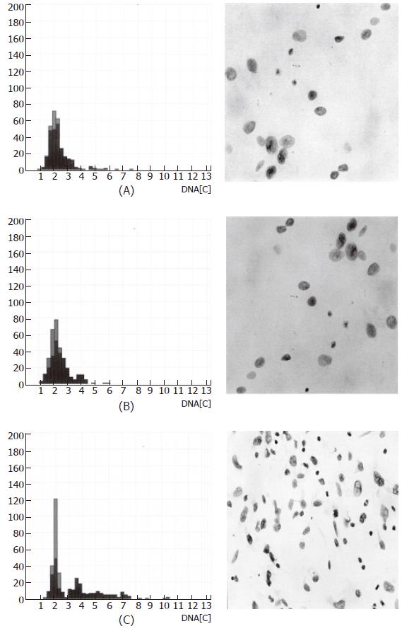
Figure 2 Image cytometric DNA analysis: histogram and nuclear morphology.
(A) achalasia; (B) peritumoral tissue (esophageal carcinoma); (C) tumor center (esophageal carcinoma).
- Citation: Gockel I, Kämmerer P, Brieger J, Heinrich U, Mann W, Bittinger F, Eckardt V, Junginger T. Image cytometric DNA analysis of mucosal biopsies in patients with primary achalasia. World J Gastroenterol 2006; 12(19): 3020-3025
- URL: https://www.wjgnet.com/1007-9327/full/v12/i19/3020.htm
- DOI: https://dx.doi.org/10.3748/wjg.v12.i19.3020


