Copyright
©2006 Baishideng Publishing Group Co.
World J Gastroenterol. May 21, 2006; 12(19): 2969-2978
Published online May 21, 2006. doi: 10.3748/wjg.v12.i19.2969
Published online May 21, 2006. doi: 10.3748/wjg.v12.i19.2969
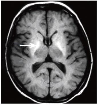
Figure 1 T1-weighted brain MR image through the basal ganglia acquired at 1.
5T, showing bilateral globus pallidus (arrow) hyperintensity in a patient with cirrhosis of the liver.
- Citation: Grover VB, Dresner MA, Forton DM, Counsell S, Larkman DJ, Patel N, Thomas HC, Taylor-Robinson SD. Current and future applications of magnetic resonance imaging and spectroscopy of the brain in hepatic encephalopathy. World J Gastroenterol 2006; 12(19): 2969-2978
- URL: https://www.wjgnet.com/1007-9327/full/v12/i19/2969.htm
- DOI: https://dx.doi.org/10.3748/wjg.v12.i19.2969


