Copyright
©2006 Baishideng Publishing Group Co.
World J Gastroenterol. May 14, 2006; 12(18): 2846-2857
Published online May 14, 2006. doi: 10.3748/wjg.v12.i18.2846
Published online May 14, 2006. doi: 10.3748/wjg.v12.i18.2846
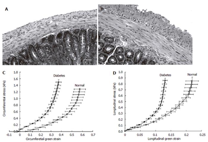
Figure 4 The micro-photographs show the normal (A) and 4 wk STZ-induced diabetic (B) duodenal histological sections.
It clearly demonstrates that the muscle and submucosa layers in the diabetic duodenum became much thicker than in the normal duodenum. The bar is 100 μm. Correspondingly, the circumferential (C) and longitudinal (D) stress-strain relation curves of the duodenum in the STZ-induced diabetic rats shifted to the left indicating the wall became stiffer.
- Citation: Zhao JB, Frøkjær JB, Drewes AM, Ejskjaer N. Upper gastrointestinal sensory-motor dysfunction in diabetes mellitus. World J Gastroenterol 2006; 12(18): 2846-2857
- URL: https://www.wjgnet.com/1007-9327/full/v12/i18/2846.htm
- DOI: https://dx.doi.org/10.3748/wjg.v12.i18.2846


