Copyright
©2006 Baishideng Publishing Group Co.
World J Gastroenterol. May 7, 2006; 12(17): 2749-2755
Published online May 7, 2006. doi: 10.3748/wjg.v12.i17.2749
Published online May 7, 2006. doi: 10.3748/wjg.v12.i17.2749
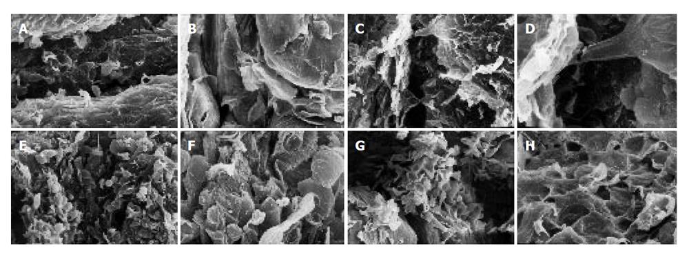
Figure 3 SEM of forestomach of mice.
A: Control mice showing grooves and ridges X 200; B: Control mice showing tongue-shaped squamous epithelium X 600; C: Mice treated with only AAILE showing grooves and ridges X 200; D: Mice treated with only AAILE showing squamous epithelial cells X 600; E: Mice received only B(a)P showing ruptured surface X 200; F: Mice received only B(a)P showing certain rounded structures in addition to tongue-shaped squamous epithelial cells X 600; G: Mice received AAILE along with B(a)P instillations showing surface rupturing X 200; H: Mice received AAILE along with B(a)P instillations showing tongue shaped squamous epithelial cells X 600.
- Citation: Gangar SC, Sandhir R, Rai DV, Koul A. Modulatory effects of Azadirachta indica on benzo(a)pyrene-induced forestomach tumorigenesis in mice. World J Gastroenterol 2006; 12(17): 2749-2755
- URL: https://www.wjgnet.com/1007-9327/full/v12/i17/2749.htm
- DOI: https://dx.doi.org/10.3748/wjg.v12.i17.2749


