Copyright
©2006 Baishideng Publishing Group Co.
World J Gastroenterol. May 7, 2006; 12(17): 2749-2755
Published online May 7, 2006. doi: 10.3748/wjg.v12.i17.2749
Published online May 7, 2006. doi: 10.3748/wjg.v12.i17.2749
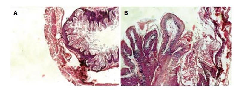
Figure 2 Histo-micrograph of forestomach.
A: Control mice showing squamous mucosa, sub-mucosa and muscular layers X 20; B: Papillary projections, disrupted submucosa and muscular layers X 20.
- Citation: Gangar SC, Sandhir R, Rai DV, Koul A. Modulatory effects of Azadirachta indica on benzo(a)pyrene-induced forestomach tumorigenesis in mice. World J Gastroenterol 2006; 12(17): 2749-2755
- URL: https://www.wjgnet.com/1007-9327/full/v12/i17/2749.htm
- DOI: https://dx.doi.org/10.3748/wjg.v12.i17.2749


