Copyright
©2006 Baishideng Publishing Group Co.
World J Gastroenterol. May 7, 2006; 12(17): 2641-2655
Published online May 7, 2006. doi: 10.3748/wjg.v12.i17.2641
Published online May 7, 2006. doi: 10.3748/wjg.v12.i17.2641
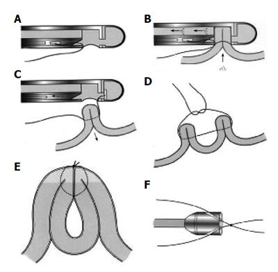
Figure 2 A: Drawing of the suturing device which is equipped with a vacuum chamber and a hollow needle in which there is a suture attached to a tag; B: Tissue is drawn into the chamber by suction.
The needle passes through the tissue and the tag is captured in the distal chamber; C: The suction is then released; D: The procedure is repeated on an adjacent piece of tissue; E: On tightening the knot, the pieces of tissues are approximated; F: The knot, tied outside the animal, is advanced by a knot pusher which is attached to the tip of the endoscope.
- Citation: Iqbal A, Salinas V, Filipi CJ. Endoscopic therapies of gastroesophageal reflux disease. World J Gastroenterol 2006; 12(17): 2641-2655
- URL: https://www.wjgnet.com/1007-9327/full/v12/i17/2641.htm
- DOI: https://dx.doi.org/10.3748/wjg.v12.i17.2641


