Copyright
©2006 Baishideng Publishing Group Co.
World J Gastroenterol. Apr 28, 2006; 12(16): 2629-2632
Published online Apr 28, 2006. doi: 10.3748/wjg.v12.i16.2629
Published online Apr 28, 2006. doi: 10.3748/wjg.v12.i16.2629
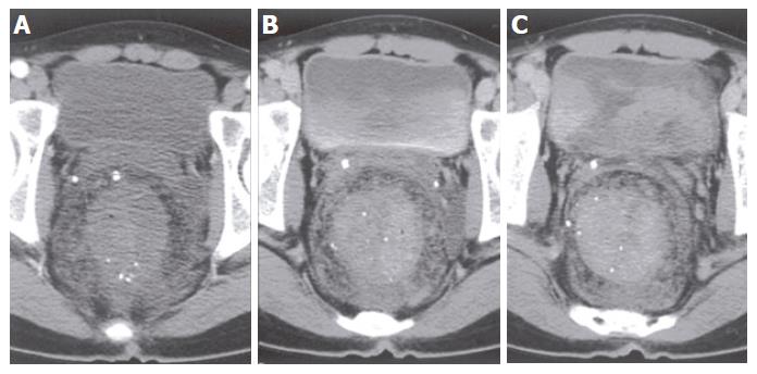
Figure 2 Abdomen-pelvis triphasic computed tomography showing an arterial phase image of mucosal enhacement (A) venous (B) and delayed(C) phase images of delayed diffuse wall enhancement.
- Citation: Han JH, Jeon WJ, Chae HB, Park SM, Youn SJ, Kim SH, Bae IH, Lee SJ. A case of idiopathic colonic varices: A rare cause of hematochezia misconceived as tumor. World J Gastroenterol 2006; 12(16): 2629-2632
- URL: https://www.wjgnet.com/1007-9327/full/v12/i16/2629.htm
- DOI: https://dx.doi.org/10.3748/wjg.v12.i16.2629


