Copyright
©2006 Baishideng Publishing Group Co.
World J Gastroenterol. Apr 28, 2006; 12(16): 2615-2619
Published online Apr 28, 2006. doi: 10.3748/wjg.v12.i16.2615
Published online Apr 28, 2006. doi: 10.3748/wjg.v12.i16.2615
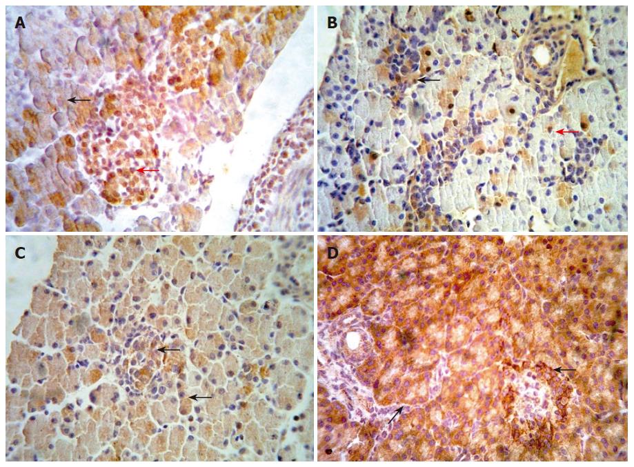
Figure 2 (A-D) Representative immunohistochemical results of β-catenin, APC and cyclin D1 proteins in embryonic pancreas of E14.
5-E18.5 and adult rat. Immunohistochemical staining was performed using an anti-β-catenin APC and cyclin D1antibodies respectively. Slides were detected at ×400 magnification under a light microscope. A: showed staining of β-catenin at cytoplasm (black arrow) and nucleus (red row) in embryonic pancreas of E18.5. B: showed staining of cyclin D1 within cytoplasm (black arrow) and/or nucleus (red row) in embryonic pancreas of E16.5. C: showed staining of APC within cytoplasm (black arrow) in embryonic pancreas of E18.5. D: showed staining of APC within cytoplasm (black arrow) in adult rat pancreas.
- Citation: Wang QM, Zhang Y, Yang KM, Zhou HY, Yang HJ. Wnt/β-catenin signaling pathway is active in pancreatic development of rat embryo. World J Gastroenterol 2006; 12(16): 2615-2619
- URL: https://www.wjgnet.com/1007-9327/full/v12/i16/2615.htm
- DOI: https://dx.doi.org/10.3748/wjg.v12.i16.2615


