Copyright
©2006 Baishideng Publishing Group Co.
World J Gastroenterol. Apr 28, 2006; 12(16): 2510-2516
Published online Apr 28, 2006. doi: 10.3748/wjg.v12.i16.2510
Published online Apr 28, 2006. doi: 10.3748/wjg.v12.i16.2510
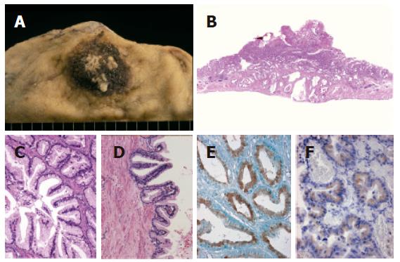
Figure 1 Extremely well-differentiated adenocarcinoma of the stomach, gastric-type (case 4).
A: Macroscopic view showing a polypoid lesion with an irregular surface; B: cancer invasion of the whole thickness of the gastric wall (low-power view); C: carcinoma mimicking the normal gastric foveolar epithelium with basally located small nuclei (hyperchromatic nuclei) and abundant mucin; D: papillary projections occasionally seen in the carcinomatous glands; E: diffuse positive staining of human gastric mucin in carcinomatous glands; and F: focally positive staining of MUC6 in carcinomatous glands.
- Citation: Yao T, Utsunomiya T, Oya M, Nishiyama K, Tsuneyoshi M. Extremely well-differentiated adenocarcinoma of the stomach: Clinicopathological and immunohistochemical features. World J Gastroenterol 2006; 12(16): 2510-2516
- URL: https://www.wjgnet.com/1007-9327/full/v12/i16/2510.htm
- DOI: https://dx.doi.org/10.3748/wjg.v12.i16.2510


