Copyright
©2006 Baishideng Publishing Group Co.
World J Gastroenterol. Apr 14, 2006; 12(14): 2209-2216
Published online Apr 14, 2006. doi: 10.3748/wjg.v12.i14.2209
Published online Apr 14, 2006. doi: 10.3748/wjg.v12.i14.2209
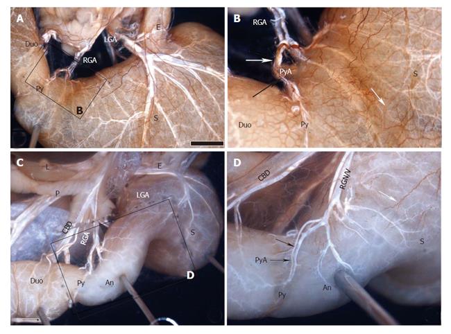
Figure 5 Two cases showing innervation of the pyloric region (Py) in Suncus murinus by whole-mount immunostaining.
High magnifications of the boxed areas in A and C are shown in B and D. White arrows show the nerves of Latarjet intended for the antro-pyloric region. Black arrows indicate the nerves arising from the right gastric artery (RGA), running along the pyloric artery (PyA), and reaching the pyloric region. An, pyloric antrum; CBD, common bile duct; Duo, duodenum; E, esophagus; L, liver; LGA, left gastric artery; P, pancreas; RGA/V, right gastric artery/vein; S, stomach. Scale bar = 2 mm in A and B.
- Citation: Yi SQ, Ru F, Ohta T, Terayama H, Naito M, Hayashi S, Buhe S, Yi N, Miyaki T, Tanaka S, Itoh M. Surgical anatomy of the innervation of pylorus in human and Suncus murinus, in relation to surgical technique for pylorus-preserving pancreaticoduodenectomy. World J Gastroenterol 2006; 12(14): 2209-2216
- URL: https://www.wjgnet.com/1007-9327/full/v12/i14/2209.htm
- DOI: https://dx.doi.org/10.3748/wjg.v12.i14.2209


