Copyright
©2006 Baishideng Publishing Group Co.
World J Gastroenterol. Apr 14, 2006; 12(14): 2209-2216
Published online Apr 14, 2006. doi: 10.3748/wjg.v12.i14.2209
Published online Apr 14, 2006. doi: 10.3748/wjg.v12.i14.2209
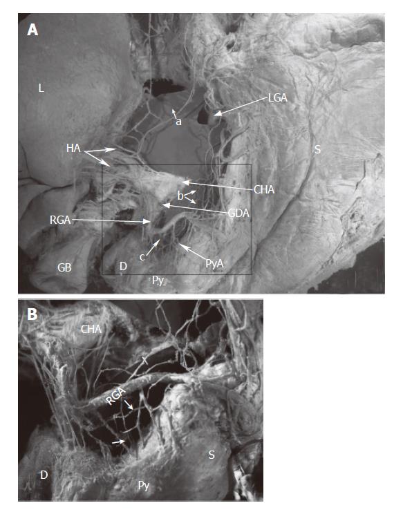
Figure 1 The innervation of the pyloric region from the view of the superior part of the pylorus, its schematic shows in Figure 3A.
Hepatic divisions (a) join in the anterior hepatic plexus in the proper hepatic artery or the left/right hepatic artery (HA). The nerves ran along the right gastric artery (RGA) and its branches (PyA), and reached the pyloric region (Py) (c). The nerves (b) of Latarjet ran along the lesser curvature or the branches of the left gastric artery (LGA), intended for the antro-pyloric region. B is an enlargement of the box in A., the arrows show the nerves innervating the pyloric region from the right gastric artery. CHA, common hepatic artery; D, duodenum; E, esophagus; GB, gallbladder; GDA, gastroduodenal artery; L, liver; S, stomach.
- Citation: Yi SQ, Ru F, Ohta T, Terayama H, Naito M, Hayashi S, Buhe S, Yi N, Miyaki T, Tanaka S, Itoh M. Surgical anatomy of the innervation of pylorus in human and Suncus murinus, in relation to surgical technique for pylorus-preserving pancreaticoduodenectomy. World J Gastroenterol 2006; 12(14): 2209-2216
- URL: https://www.wjgnet.com/1007-9327/full/v12/i14/2209.htm
- DOI: https://dx.doi.org/10.3748/wjg.v12.i14.2209


