Copyright
©2006 Baishideng Publishing Group Co.
World J Gastroenterol. Apr 7, 2006; 12(13): 2130-2132
Published online Apr 7, 2006. doi: 10.3748/wjg.v12.i13.2130
Published online Apr 7, 2006. doi: 10.3748/wjg.v12.i13.2130
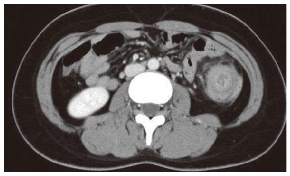
Figure 3 Computed tomography shows the cystic lesion in the descending colon, occupying the whole lumen, and also shows multicentric target sign and sausage-shaped inhomogeneous soft-tissue mass.
- Citation: Kim TO, Lee JH, Kim GH, Heo J, Kang DH, Song GA, Cho M. Adult intussusception caused by cystic lymphangioma of the colon: A rare case report. World J Gastroenterol 2006; 12(13): 2130-2132
- URL: https://www.wjgnet.com/1007-9327/full/v12/i13/2130.htm
- DOI: https://dx.doi.org/10.3748/wjg.v12.i13.2130


