Copyright
©2006 Baishideng Publishing Group Co.
World J Gastroenterol. Apr 7, 2006; 12(13): 2016-2023
Published online Apr 7, 2006. doi: 10.3748/wjg.v12.i13.2016
Published online Apr 7, 2006. doi: 10.3748/wjg.v12.i13.2016
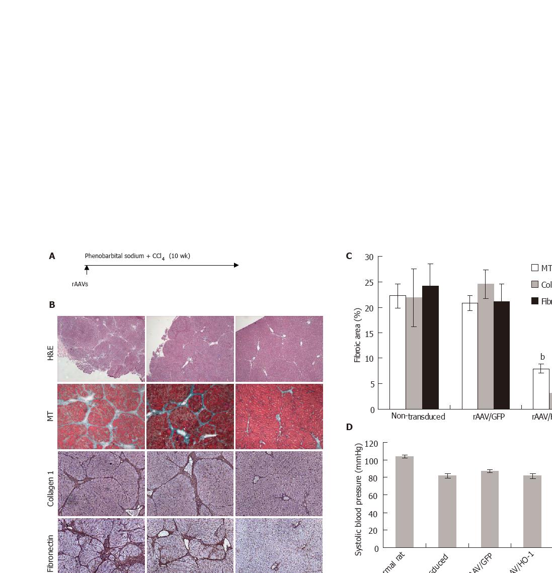
Figure 2 A: The model to induce micronodular cirrhosis in LEW rat.
The rAAVs were injected through the portal vein shortly before the induction; B: The histology and immunohistochemistry of the livers with or without treatment at the 10th week (H-E, original magnification ×40; MT: Masson’s trichrome stiaining, the fibrotic elements were stained in green, original 100×; collagen 1 and fibronectin staining, original magnification ×100); C: The measurement of fibrotic area in the livers at the 10th week by the computer software as described in Materials & Methods. Data were shown as mean±SE, n = 5, bP < 0.001; D and E: The systolic and portal pressure of rats after induction of liver cirrhosis with or without treatments. The data were shown as mean±SE, n = 10, dP < 0.001.
- Citation: Tsui TY, Lau CK, Ma J, Glockzin G, Obed A, Schlitt HJ, Fan ST. Adeno-associated virus-mediated heme oxygenase-1 gene transfer suppresses the progression of micronodular cirrhosis in rats. World J Gastroenterol 2006; 12(13): 2016-2023
- URL: https://www.wjgnet.com/1007-9327/full/v12/i13/2016.htm
- DOI: https://dx.doi.org/10.3748/wjg.v12.i13.2016


