Copyright
©2006 Baishideng Publishing Group Co.
World J Gastroenterol. Mar 28, 2006; 12(12): 1941-1944
Published online Mar 28, 2006. doi: 10.3748/wjg.v12.i12.1941
Published online Mar 28, 2006. doi: 10.3748/wjg.v12.i12.1941
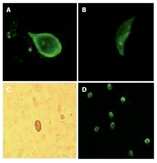
Figure 2 Confocal microscopy images (A and B) of G.
lamblia trophozoite (x1000) in duodenal bioptic sample, using acridine orange as a vital stain. A: a dorsal projection showing the peculiar tear-shaped cell with flagella. B: an unusual flank projection shows the nuclei on the back of the ventral disk. C: G. lamblia cysts (x200) microscopic images after a wet mount with Lugol’s iodine of fecal specimens. D: G. lamblia cysts (x200) in stool sample following formalin/ether enrichment of filtered samples and staining with a direct fluorescent antibody.
- Citation: Grazioli B, Matera G, Laratta C, Schipani G, Guarnieri G, Spiniello E, Imeneo M, Amorosi A, Focà A, Luzza F. Giardia lamblia infection in patients with irritable bowel syndrome and dyspepsia: A prospective study. World J Gastroenterol 2006; 12(12): 1941-1944
- URL: https://www.wjgnet.com/1007-9327/full/v12/i12/1941.htm
- DOI: https://dx.doi.org/10.3748/wjg.v12.i12.1941


