Copyright
©2006 Baishideng Publishing Group Co.
World J Gastroenterol. Mar 28, 2006; 12(12): 1933-1936
Published online Mar 28, 2006. doi: 10.3748/wjg.v12.i12.1933
Published online Mar 28, 2006. doi: 10.3748/wjg.v12.i12.1933
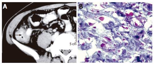
Figure 2 Contrast-enhanced CT scan of the abdomen showing the ileocecal mass with thickening of the cecal wall (A) and histological examination of the biopsies showing trophozoites of E.
histolytica (B) (H & E X 800).
- Citation: Misra SP, Misra V, Dwivedi M. Ileocecal masses in patients with amebic liver abscess: Etiology and management. World J Gastroenterol 2006; 12(12): 1933-1936
- URL: https://www.wjgnet.com/1007-9327/full/v12/i12/1933.htm
- DOI: https://dx.doi.org/10.3748/wjg.v12.i12.1933


