Copyright
©2006 Baishideng Publishing Group Co.
World J Gastroenterol. Mar 28, 2006; 12(12): 1881-1888
Published online Mar 28, 2006. doi: 10.3748/wjg.v12.i12.1881
Published online Mar 28, 2006. doi: 10.3748/wjg.v12.i12.1881
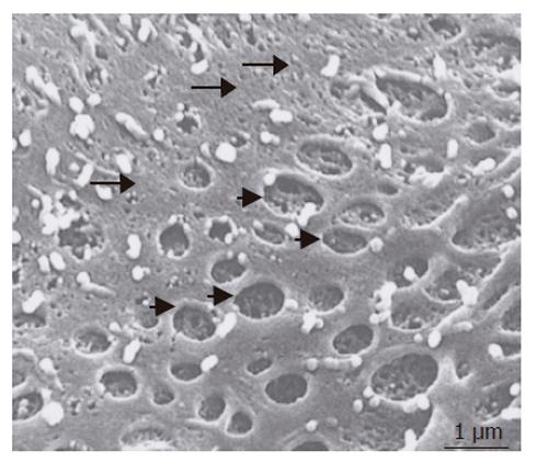
Figure 6 Scanning electron micrographs of the surface of swinholide A - treated SEC cells in the RFB culture system.
Large open pores have a fenestra-like appearance (short arrow). Small pores were detectable in the nonfenestrated area (long arrow). Scale bar: 1 μm.
- Citation: Saito M, Matsuura T, Masaki T, Maehashi H, Shimizu K, Hataba Y, Iwahori T, Suzuki T, Braet F. Reconstruction of liver organoid using a bioreactor. World J Gastroenterol 2006; 12(12): 1881-1888
- URL: https://www.wjgnet.com/1007-9327/full/v12/i12/1881.htm
- DOI: https://dx.doi.org/10.3748/wjg.v12.i12.1881


