Copyright
©2006 Baishideng Publishing Group Co.
World J Gastroenterol. Mar 21, 2006; 12(11): 1699-1705
Published online Mar 21, 2006. doi: 10.3748/wjg.v12.i11.1699
Published online Mar 21, 2006. doi: 10.3748/wjg.v12.i11.1699
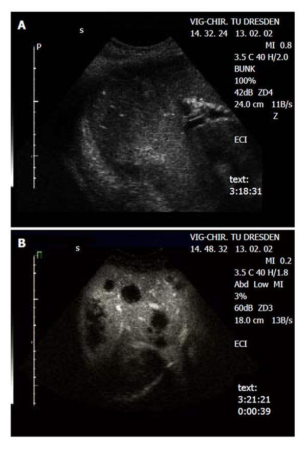
Figure 2 Detection of metastases in a patient with colorectal carcinoma.
A: Native B-mode sonography revealed 3 metastases (segment 6/7) in agreement with CT, MRI revealed 4 metastases (segment 6/7 and 4). B: Contrast-enhanced sonography identified diffuse metastatic disease in both liver lobes. The metastatic lesions are clearly delineated in the portal-venous phase as ‘black spots’, due to the lack of portal-venous blood supply.
- Citation: Dietrich CF, Kratzer W, Strobel D, Danse E, Fessl R, Bunk A, Vossas U, Hauenstein K, Koch W, Blank W, Oudkerk M, Hahn D, Greis C. Assessment of metastatic liver disease in patients with primary extrahepatic tumors by contrast-enhanced sonography versus CT and MRI. World J Gastroenterol 2006; 12(11): 1699-1705
- URL: https://www.wjgnet.com/1007-9327/full/v12/i11/1699.htm
- DOI: https://dx.doi.org/10.3748/wjg.v12.i11.1699


