Copyright
©2006 Baishideng Publishing Group Co.
World J Gastroenterol. Mar 14, 2006; 12(10): 1630-1633
Published online Mar 14, 2006. doi: 10.3748/wjg.v12.i10.1630
Published online Mar 14, 2006. doi: 10.3748/wjg.v12.i10.1630
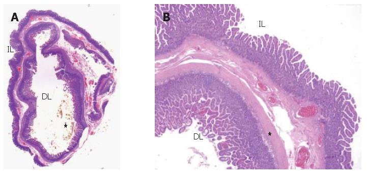
Figure 6 Mucosa, submucosa and muscle coats found in pathological ehamination.
A: Gross transversal section of the specimen “in toto”: inside it is possible to see the duplication lumen (DL) while the intestinal lumen is outside (IL). In the inner lumen there are bile plugs (*); B: Histology of the duodenal duplication. Microscopic low power view of the duplication wall (2x). The wall is composed of muscolaris propria extending into the septum between the duplicated segment and the mucosal lining. Intestinal mucosa is on both sides of the duplication, with a lot of macrophage cells (*) in the lamina propria of the internal one.
- Citation: Guarise A, Faccioli N, Ferrari M, Romano L, Parisi A, Falconi M. Duodenal duplication cyst causing severe pancreatitis: Imaging findings and pathological correlation. World J Gastroenterol 2006; 12(10): 1630-1633
- URL: https://www.wjgnet.com/1007-9327/full/v12/i10/1630.htm
- DOI: https://dx.doi.org/10.3748/wjg.v12.i10.1630


