Copyright
©2006 Baishideng Publishing Group Co.
World J Gastroenterol. Mar 14, 2006; 12(10): 1569-1576
Published online Mar 14, 2006. doi: 10.3748/wjg.v12.i10.1569
Published online Mar 14, 2006. doi: 10.3748/wjg.v12.i10.1569
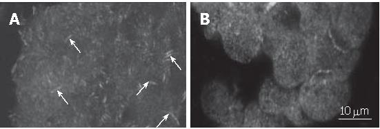
Figure 4 Immunocytochemical localisation of FAK.
DSL6A cells grown on glass cover slips were cultured for 24 h without (A) or with 10 mmol/L CHP (B). Cell staining was performed by incubation with a mouse anti-FAK specific antibody. Binding of the primary antibody was detected by an AlexaFluorTM-labelled anti-mouse antibody. Arrows: focal adhesions; Bar: 10 μm.
- Citation: Mueller C, Emmrich J, Jaster R, Braun D, Liebe S, Sparmann G. Cis-hydroxyproline-induced inhibition of pancreatic cancer cell growth is mediated by endoplasmic reticulum stress. World J Gastroenterol 2006; 12(10): 1569-1576
- URL: https://www.wjgnet.com/1007-9327/full/v12/i10/1569.htm
- DOI: https://dx.doi.org/10.3748/wjg.v12.i10.1569


