Copyright
©2005 Baishideng Publishing Group Inc.
World J Gastroenterol. Mar 7, 2005; 11(9): 1403-1409
Published online Mar 7, 2005. doi: 10.3748/wjg.v11.i9.1403
Published online Mar 7, 2005. doi: 10.3748/wjg.v11.i9.1403
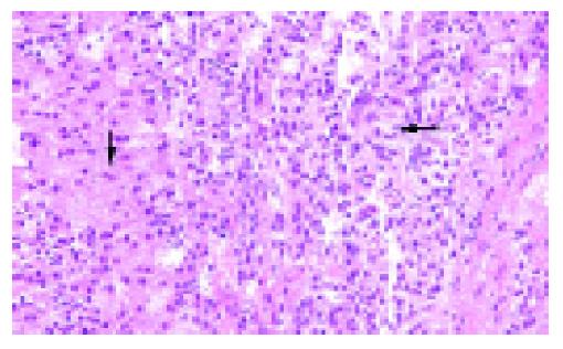
Figure 8 Metastatic tumor in liver (case 1) showing small tumor cells (straight arrow) compared to the normal liver cells with abundant cytoplasm (curved arrow) (HE, original magnification ×200).
- Citation: Huang HL, Shih SC, Chang WH, Wang TE, Chen MJ, Chan YJ. Solid-pseudopapillary tumor of the pancreas: Clinical experience and literature review. World J Gastroenterol 2005; 11(9): 1403-1409
- URL: https://www.wjgnet.com/1007-9327/full/v11/i9/1403.htm
- DOI: https://dx.doi.org/10.3748/wjg.v11.i9.1403


