Copyright
©2005 Baishideng Publishing Group Inc.
World J Gastroenterol. Mar 7, 2005; 11(9): 1345-1350
Published online Mar 7, 2005. doi: 10.3748/wjg.v11.i9.1345
Published online Mar 7, 2005. doi: 10.3748/wjg.v11.i9.1345
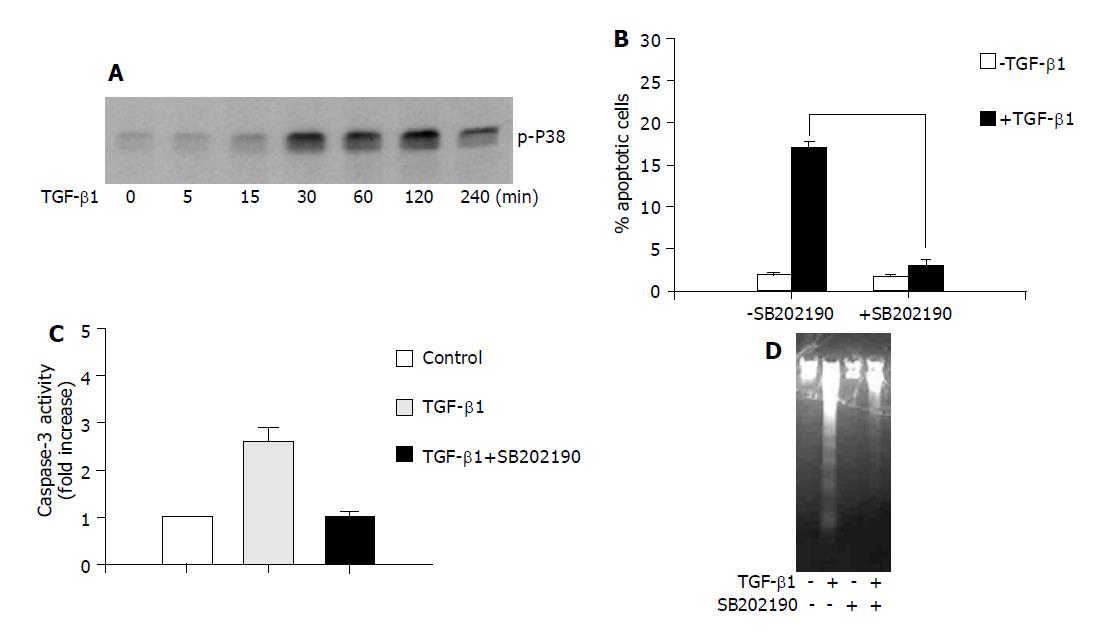
Figure 1 The role of p38 in TGF-β1-induced apoptosis in AML-12 hepatocytes.
A: AML-12 cells were treated with TGF-β1 (5 ng/mL) for the indicated time, and the phosphorylation of p38 was examined with anti phospho-p38 antibody; B, C and D: Cells were treated with TGF-β1 (5 ng/mL) for 24 h in the presence or absence of SB202190 (10 μmol/L). Apoptotic rate was determined by measuring DNA content with flow cytometry analysis (B); the caspase-3 activity (C); and DNA fragmentation assay (D).
- Citation: Guo LX, Xie H. Differential phosphorylation of p38 induced by apoptotic and anti-apoptotic stimuli in murine hepatocytes. World J Gastroenterol 2005; 11(9): 1345-1350
- URL: https://www.wjgnet.com/1007-9327/full/v11/i9/1345.htm
- DOI: https://dx.doi.org/10.3748/wjg.v11.i9.1345


