Copyright
©2005 Baishideng Publishing Group Inc.
World J Gastroenterol. Mar 7, 2005; 11(9): 1317-1323
Published online Mar 7, 2005. doi: 10.3748/wjg.v11.i9.1317
Published online Mar 7, 2005. doi: 10.3748/wjg.v11.i9.1317
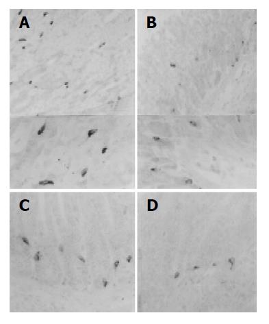
Figure 2 Somatostatin-IR cells in the fundus (A, B) and pylorus (C, D) of non-implanted sham (A, B) and 3LL-implanted group (B, D).
In 3LL-implanted group, somatostatin-IR cells dramatically decreased in both regions of stomach and cells having relative narrow cytoplasm and degranulation were more numerously detected. PAP methods, scale bar = 40 μm.
- Citation: Ku SK, Lee HS, Byun JS, Seo BI, Lee JH. Changes of the gastric endocrine cells in the C57BL/6 mouse after implantation of murine lung carcinoma: An immunohistochemical quantitative study. World J Gastroenterol 2005; 11(9): 1317-1323
- URL: https://www.wjgnet.com/1007-9327/full/v11/i9/1317.htm
- DOI: https://dx.doi.org/10.3748/wjg.v11.i9.1317


