Copyright
©2005 Baishideng Publishing Group Inc.
World J Gastroenterol. Mar 7, 2005; 11(9): 1303-1316
Published online Mar 7, 2005. doi: 10.3748/wjg.v11.i9.1303
Published online Mar 7, 2005. doi: 10.3748/wjg.v11.i9.1303
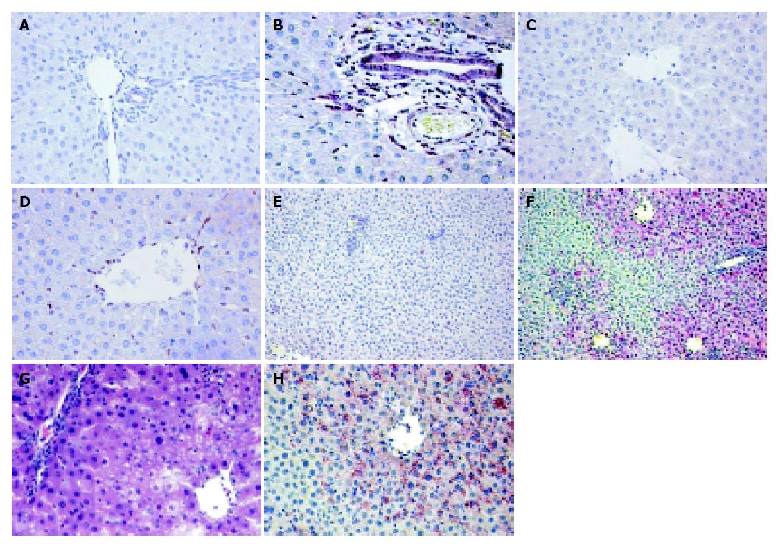
Figure 8 Hsp70 protein localization in rat livers after WI/R (paraffin-embedded sections).
Hsp70 staining of paraffin-embedded liver tissues after partial WI/R. Up to 2 h of reperfusion Hsp70 was located in sinusoidal cells and cholangiocytes, connective cells, endothelial cells of portal areas only in the I-lobes (I-lobes after 1 h/2 h WI/R: B+D), while Hsp70 was not detectable in the C-lobes at these time points (C-lobes after 1 h/2 h WI/R: A+C). Parenchymal Hsp70 staining was only noticed after 6 h reperfusion (C-lobe: E; I-lobe: F+H).Comparison of perivenular vacuolization (HE staining: G) and Hsp70 protein expression in the affected hepatocytes after 1 h/6 h WI/R (H).
- Citation: Fallsehr C, Zapletal C, Kremer M, Demir R, von Knebel Doeberitz M, Klar E. Identification of differentially expressed genes after partial rat liver ischemia/reperfusion by suppression subtractive hybridization. World J Gastroenterol 2005; 11(9): 1303-1316
- URL: https://www.wjgnet.com/1007-9327/full/v11/i9/1303.htm
- DOI: https://dx.doi.org/10.3748/wjg.v11.i9.1303


