Copyright
©2005 Baishideng Publishing Group Inc.
World J Gastroenterol. Feb 28, 2005; 11(8): 1179-1181
Published online Feb 28, 2005. doi: 10.3748/wjg.v11.i8.1179
Published online Feb 28, 2005. doi: 10.3748/wjg.v11.i8.1179
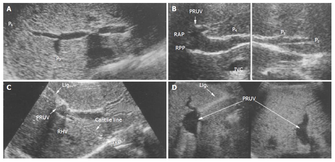
Figure 1 Persistent right umbilical vein shown on ultrasonogram.
A: Right posterior portal branch originating from the main portal vein at the portal hepatis; B: Anterior portal branch and P4 branch of the portal vein originating from the umbilical portion; C, D: Persistent right umbilical vein on the Cantlie line; PRUV: Persistent right umbilical vein; RAP: right anterior portal vein; RPP: right posterior portal vein; Lig: Ligamentum teres; RHV: Right hepatic vein.
- Citation: Nakanishi S, Shiraki K, Yamamoto K, Koyama M, Nakano T. An anomaly in persistent right umbilical vein of portal vein diagnosed by ultrasonography. World J Gastroenterol 2005; 11(8): 1179-1181
- URL: https://www.wjgnet.com/1007-9327/full/v11/i8/1179.htm
- DOI: https://dx.doi.org/10.3748/wjg.v11.i8.1179


