Copyright
©2005 Baishideng Publishing Group Inc.
World J Gastroenterol. Feb 28, 2005; 11(8): 1141-1148
Published online Feb 28, 2005. doi: 10.3748/wjg.v11.i8.1141
Published online Feb 28, 2005. doi: 10.3748/wjg.v11.i8.1141
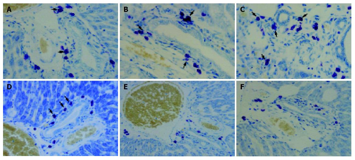
Figure 5 Toluidine blue-stained liver tissue of only CCl4- or CCl4 with silymarin-treated groups.
In only CCl4-treated group - A: At 4 wk, specific toluidine blue-stained mast cells (arrowhead) were detected in periportal region; B: At 8 wk, the number of mast cell increased; C: At 12 wk, mast cells increased along the thick collagenous septa. In CCl4 with silymarin-treated group - D: Positive reaction for toluidine blue (arrowhead) was slightly detected at 4 wk; E: At 8 wk, α-SMA-positive cells were lower than that of CCl4-only-treated group; F: At 12 wk, positive expression of α-SMA was shown in the same pattern as with the result of CCl4-treated group at 8 wk. Toluidine blue stain, A-F. Original magnification: ×132, A-F.
- Citation: Jeong DH, Lee GP, Jeong WI, Do SH, Yang HJ, Yuan DW, Park HY, Kim KJ, Jeong KS. Alterations of mast cells and TGF-β1 on the silymarin treatment for CCl4-induced hepatic fibrosis. World J Gastroenterol 2005; 11(8): 1141-1148
- URL: https://www.wjgnet.com/1007-9327/full/v11/i8/1141.htm
- DOI: https://dx.doi.org/10.3748/wjg.v11.i8.1141


